ILLUSTRATING
THE GERM-CELLS OF FISHES AND THEIR MIGRATIONS INTO THE
EMBRYONIC BODY.
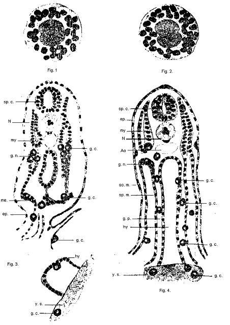
Fig. 1. - Primary germ-cell of a small dog-fish, Pristiurus
melanestomus. The cytoplasm is
glossy
in character, and contains a large number of (blackened) yolk-plates. The
nucleus exhibits
duplication. i.e., autonomy of paternal and maternal portions.
Fig. 2.-A similar primary germ-cell from the skate, Raja batis.
Fig. 3.-The conditions seen in two transverse sections of a 4 1/2 mm.
embryo of Pristiurus. The
lettering is as follows ; sp.c., spinal cord; n., notochord; my.,
myotome; g. n., germinal nidus;
me.,
mesoblast; ep., epiblast; hy., hypoblast; y. s., yolk-sac: g. c.,
germ-cell.
Fig. 4. - A diagrammatic section of an early skate-embryo. To illustrate
migrations of the
Germ-cells along the germinal path, g.p., and showing germ-cells in
various abnormal positions.
The
lettering as in Fig. 3. excepting so. m. somatic mesoblast; sp. m. splanchuic mesoblast;
ao.,
aorta.
58a fig.